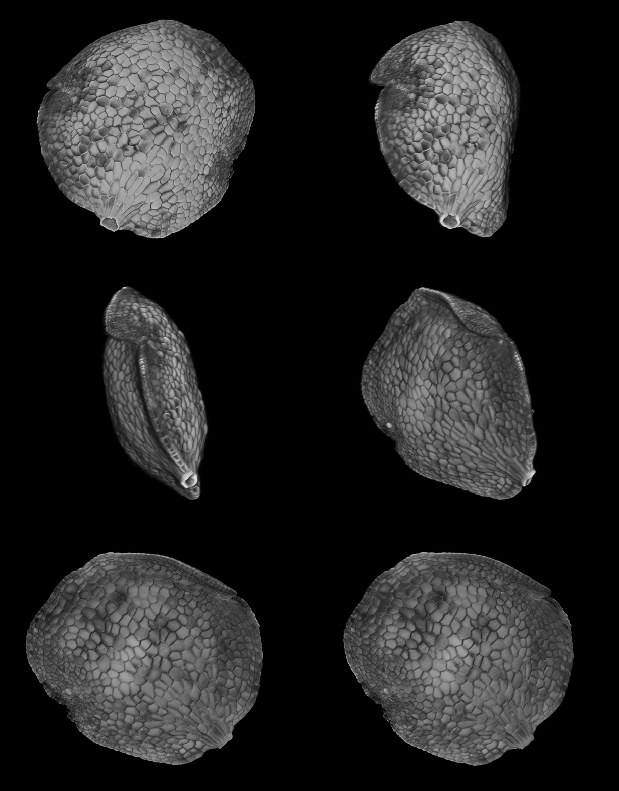
Plant scientists at Cambridge and Oxford have come up with a clever method that can produce full three-dimensional (3D) images of plant tissues using a laser scanning confocal microscope.
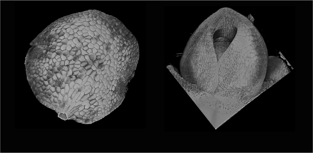
3D reconstruction of Marchantia using the new Flip-Flap protocol.
The simple imaging method uses innovative ideas and involved designing new tools to enable imaging of thick plant samples, which are challenging to image using traditional methods.
Plants have a complex 3D anatomy and visualising how they grow and develop in 3D is vital for researchers to be able to understand how plant tissues like roots and meristems grow and develop.
There are a number of imaging techniques for imaging surfaces in 3D, but getting an accurate view deeper into the tissue, especially for larger samples over 100 µm, is challenging, even when using advanced scanning electron microscopes (SEM) or X-ray computed tomography.
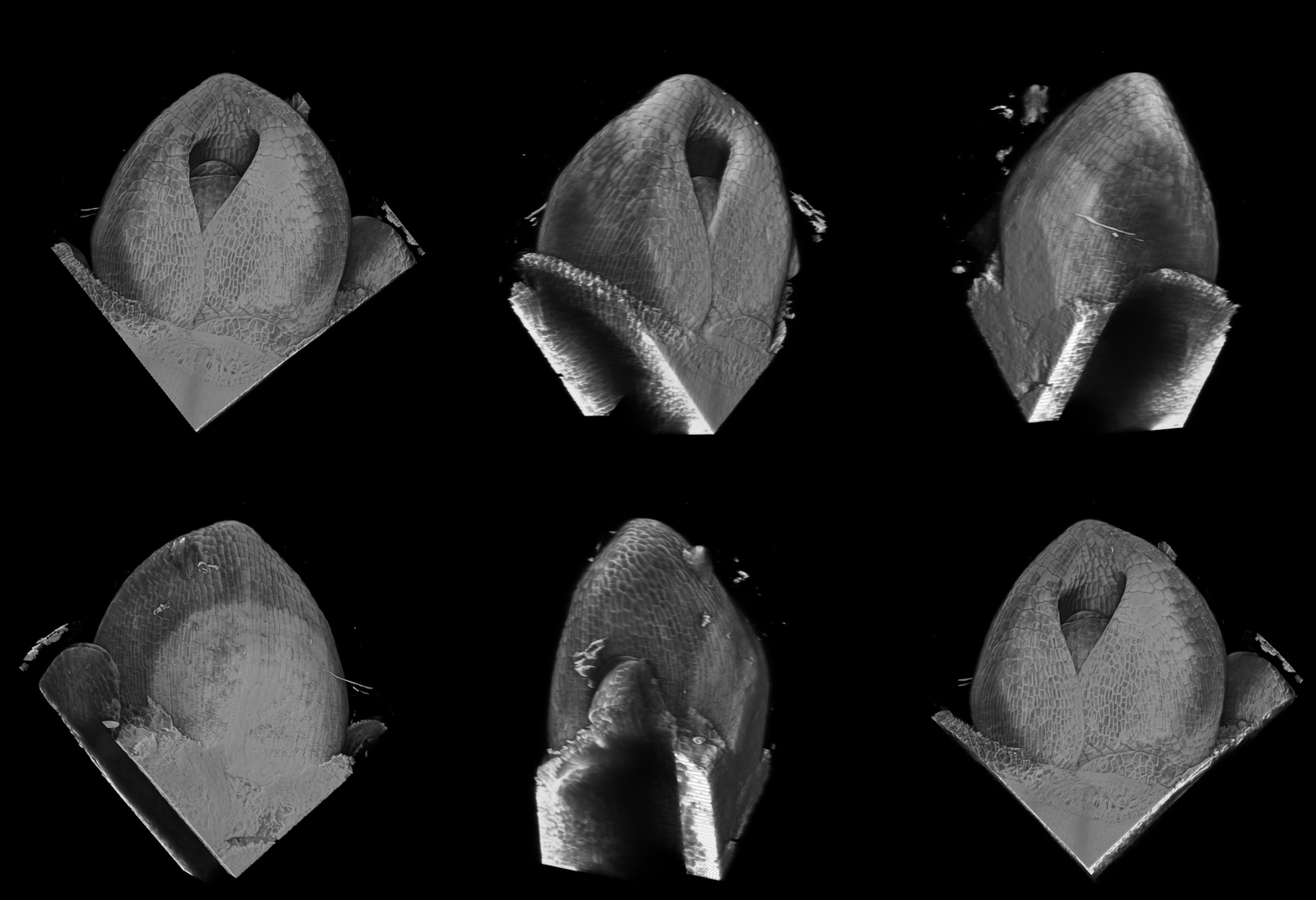
Dr Leo Serra, Research Associate in the Robinson Group at the Sainsbury Laboratory Cambridge University (SLCU), and Dr Sovanna Tan, Research Associate in Professor Jane Langdale’s group in the Department of Plant Sciences at the University of Oxford, co-authored the Flip-Flap protocol published in Plants’ Special Issue Imaging Tools for the Plant Sciences, which demonstrates Flip-Flap’s capability by producing 3D reconstructions of barley meristem and Marchantia gemma.
“An important step towards a better understanding of complexity in plants is being able to acquire 3D images of entire organs,” Dr Serra said. "However, 3D imaging of intact plant samples is not always simple and often requires expensive and complex processes. The aim of Flip-Flap is to give access to 3D imaging to all researchers and to make the process straight forward and accessible without requiring specialist techniques or advanced equipment."
3D reconstruction of barley meristem using the new Flip-Flap protocol.
Dr Sarah Robinson’s research group at SLCU is not new to adapting and inventing new innovative methods to use microscopes to capture images not previously possible.
“We are seeing some very exciting advances in microscopy at the moment,” Dr Robinson said. “These technological advances are helping us to see previously unseen processes in plants and gain a better understanding of the developmental processes and observed characteristics. The implications are huge. While we can see with the naked eye the grains and fruit of harvests, their final characteristics start at the microscopic level where stem cells first start to differentiate and patterns form in developing plant organs. Being able to dive deep into the tissues of these developing organs is giving us insights never before possible.”
Reference
Serra L, Tan S, Robinson S, Langdale JA. Flip-Flap: A Simple Dual-View Imaging Method for 3D Reconstruction of Thick Plant Samples. Plants. 2022; 11(4):506. https://doi.org/10.3390/plants11040506
Supplementary Materials
The following supporting information can be downloaded at: https://www.mdpi.com/article/10.3390/plants11040506/s1, Figure S1: Effects of slight x/y movements during Flip-Flap can be corrected by rotation, Video S1: Visualisation of 3D segmentation after sample stitching.

