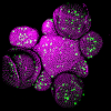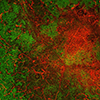
New research reveals the science behind a silver lining – SLCU researchers and CU Botanic Garden staff combine forces to discover the how and why behind the Saxifraga’s silver-white crust.
This collaboration arose from a desire to test cutting edge microscopy techniques on plants beyond those usually used in SLCU plant research. “I needed test subjects for new, state-of-the art microscopes which allowed me to look inside plant parts, cells, and even parts of cells and do chemical imaging”, explains Dr Raymond Wightman, SLCU’s Microscopy Core Facility Manager. “We knew everyday laboratory plants - such as Arabidopsis thaliana, the plant science world’s equivalent of the fruit fly - worked fine but did not know how useful the equipment would be beyond using these. This is where working in the grounds of the Botanic Garden and having the Garden’s wonderful collection of plants is so crucial to plant science research.”
Paul Aston, Alpine and Woodland Supervisor at the Botanic Garden and his assistant, Simon Wallis, introduced Ray to the encrusted Saxifraga, which gets its name from crusts that forms on leaf secretory structures called hydathodes. The study focused on Saxifraga cochlearis because of the way the hydathodes are arranged in a regular fashion along the length of the leaf. On a single leaf you can observe the hydathodes becoming more developed as you move progressively to the leaf tip, starting off as a group of small cells and ending as a volcano-type structure that spews out the crust.
“Their tough, leathery and crusty leaves provided a challenge for the process of cryo-scanning electron microscopy, a type of microscopy that relies on low temperatures of minus 150 degrees Celsius, but the equipment worked remarkably well and we were fortunate to come up with amazing results,” explains Raymond. “While opening up and looking inside the leaves, during a process called cryo-fracture, we realised that we were seeing things that we did not recognise from the text books. The chemical imaging also revealed other things (such as the precise arrangement of molecules in the crust) that had not been discovered before.
This latest discovery into how Saxifraga produce and lay down their silvery-white crust is very exciting and has been made possible thanks to new microscope technologies along with the availability of the wild plant material, held and looked after at the Botanic Garden. Because of this we now have a completely new insight into the biology of this wonderful genus which may be able to help us develop new biomaterials for pharmaceuticals and engineering applications.”
This study highlights the possibilities of combining the collections and expertise within the Botanic Garden with cutting edge research techniques at SLCU. Paul Aston says “Raymond has been great fun to work with and it’s very rewarding for us to see the results of this partnership of the Garden and Laboratory working together.” Raymond says, “Paul and Simon were always on hand to direct me to the best plants to study and to help figure out what we were looking at.”
The results of the study will not only satisfy the curiosity of plant lovers but also demonstrate how plant science is at a pioneering stage. Raymond explains: “Modern microscopy techniques are now no longer confining plant scientists to a small number of ‘model’ plants grown in the laboratory environment. This really shows how we can leap out in to the unknown and observe things that were not possible for plant scientists just two years ago. This is just the very first part of our work on Saxifraga biology. It is all very exciting so watch this space!”
The online journal article can be read here: http://www.sciencedirect.com/science/article/pii/S0367253017332309


