A novel method that improves the images captured in light-sheet microscopy looks set to help advance the future of in vivo biological imaging.
Light-sheet microscopy is less damaging to cells and tissues than other forms of imaging like confocal and electron microscopy and allows scientists to image live biological samples. As a result, light-sheet microscopy is rapidly becoming a go-to imaging tool across the medical and life sciences.
The combination of lower photo-toxicity, better sectioning capabilities and faster image acquisition led to light-sheet microscopy being recognised as “Method of the Year” by Nature Methods in 2014. Less photo-toxicity also allows imaging of living samples over longer periods of time.
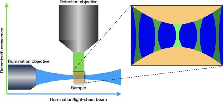
While the captured images are sharp in the centre, the quality of the image decreases towards the edges and becomes blurrier. This is mostly caused by the light sheet excitation itself. Existing deconvolution software typically do not take into account the special characteristics of light sheet microscopy.
A Cambridge collaboration led by Dr Leila Muresan has come up with a novel method for performing deconvolution for light-sheet microscopy, bringing together scientists from the Cambridge Advanced Imaging Centre (CAIC), MRC Laboratory of Molecular Biology, Department of Applied Mathematics and Theoretical Physics, and Sainsbury Laboratory (SLCU).
The method relies on advanced optimisation algorithms adapted to a new theoretical image formation model. The results show the new method outperforms existing approaches to deconvolve light-sheet microscopy data.
The experimental part of testing the method was undertaken at the Sainsbury Laboratory using a new light sheet system designed for rapid fluorescence imaging of whole roots, seedlings and plant organs, which was built by and is maintained by Dr Martin Lenz as part of a collaboration between SLCU and CAIC. Both live and cleared samples are imaged vertically in a bespoke imaging chamber containing two illumination objectives and two imaging objectives. The system allows imaging of both sides of a sample concurrently and a rotating sample holder permits multi-angle views of the sample. The system supports all the major fluorophores plus FRET-based biosensors.
The next step for the collaboration is to optimise the technique and create a user-friendly tool tailored to applied life sciences research.
Reference
Bogdan Toader, Jérôme Boulanger, Yury Korolev, Martin O. Lenz, James Manton, Carola-Bibiane Schönlieb & Leila Mureşan. (2022) Image Reconstruction in Light-Sheet Microscopy: Spatially Varying Deconvolution and Mixed Noise. J Math Imaging Vis. https://doi.org/10.1007/s10851-022-01100-3


