I am fascinated by the process of development. My research focuses on plant development and how growth, cell division and differentiation are coordinated. I am particularly interested in how cell and organ size and shape are determined and am investigating these processes using biomechanics, modelling and genomic approaches.
How dynamic spatial patterns are generated within growing proliferating tissues is crucial to understanding development in multi-cellular organisms. However, this process is poorly understood due to difficulties in unravelling the interactions between the many processes that occur at once. My PhD investigation of this problem involved looking at the patterning of stomata in growing proliferating Arabidopsis leaves at the John Innes Centres under the supervision of Enrico Coen. I followed cell lineages using live confocal time lapse imaging to compare behaviour of wild type plants with the speechless mutant, which does not make stomata. Under the supervision of my co-supervisor Przemyslaw Prusinkiewicz I generated a descriptive model of the spch mutant that provided a framework for investigating the wild-type divisions, which provided an insight into how dynamic spatial patterns can be generated in growing proliferating tissues.
 Growth was an ever-present topic during my PhD, and this motivated me to further investigate mechanics in collaboration with experts in this field as an EMBO Research Fellow in the lab of Professor Cris Kuhlemeier at the University of Bern. It was here that I developed new method for studying biomechanics in plant development.
The mechanical properties of the cell wall and their spatial variation are the key factors controlling morphogenesis in plants. However, these properties are difficult to measure and investigating their relation to genetic regulation is particularly challenging. A major issue has been the lack of suitable methods. Traditional extensometers are not suitable for the small developing Arabidopsis tissues and provide low-resolution information. Indentation methods, by contrast, provide very high-resolution information but are restricted to measuring the surface of the epidermis perpendicular to the direction of growth. I, therefore, led the development of a method (ACME) that combines confocal imaging and mechanical manipulations, with the help of an electrical engineer M. Huflejt.
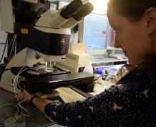
ACME robotic system to measure mechanical properties of living cells
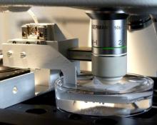 The open-source ACME (Automated confocal micro-extensometer) method (Robinson 2017 et al) was developed to measure the mechanical properties that accompany plant growth. ACME functions like a traditional extensometer, but is miniaturised, mounted on the stage of a confocal microscope, and fully automated. ACME computes the mechanical properties of samples at the cellular level based on changes in features of the tissue itself (tracked in time-lapse z-stack images) during application of a known force or deformation.
I have used the ACME system to measure the changes in the mechanical properties of the individual cells of the developing hypocotyl in an Arabidopsis seedling upon the application of the plant hormone gibberellic acid that promotes growth in the hypocotyl (Robinson et al. 2017). I also used the ACME setup to investigate whether microtubules respond to mechanical stress in Arabidopsis hypocotyls. I was able to confirm results present in the literature and to extend our knowledge of the field by directly applying known forces. I saw that microtubules reoriented in response to compression but not tension. In response to compression, the seedlings increased their growth rate (Robinson et al. 2018). We, thus, were able to demonstrate mechanical stress altering development via reorientation of microtubules.
ACME created a lot of interest from other scientists who have also been looking for better methods to capture quantitative biophysical data at cellular resolution. While it was developed to study Arabidopsis seedlings, the system can be adapted for larger samples and other imaging systems. I will continue to use the method with my SLCU research group to investigate other questions related to plant development, and work with other research groups who find this tool useful.
Read more about ACME
Publications
Alessandra Bonfanti, Euan Thomas Smithers, Matthieu Bourdon, Alex Guyon, Philip Carella, Ross Carter, Raymond Wightman, Sebastian Schornack, Henrik Jönsson, Sarah Robinson (2023) Stiffness transitions in new walls post-cell division differ between Marchantia polymorpha gemmae and Arabidopsis thaliana leaves. PNAS DOI: 10.1073/pnas.2302985120
Robinson, S. (2021), Mechanobiology of cell division in plant growth. New Phytol, 231: 559-564. https://doi.org/10.1111/nph.17369
Chiara A. Airoldi, Carlos A. Lugo, Raymond Wightman, Beverley J. Glover, Sarah Robinson* (2021) Mechanical buckling can pattern the light-diffracting cuticle of Hibiscus trionum. Cell Reports DOI: 10.1016/j.celrep.2021.109715 (*co-corresponding)
Serra L, Robinson S. (2020) Plant cell divisions: variations from the shortest symmetric path. Biochem Soc Trans. DOI:10.1042/BST20200529 (corresponding author).
Yarahmadov* T, Robinson* S, Hanemian* M, Pulver V, Kuhlemeier C. (2020) Identification of transcription factors controlling floral morphology in wild Petunia species with contrasting pollination syndromes. The Plant Journal. DOI:10.1111/tpj.14962 (co-first authors)
Bonfanti A, and Robinson S. (2020). Studying cell wall mechanics using an automated confocal micro-extensometer. Methods in Cell Biology, Academic Press. DOI: 10.1016/bs.mcb.2020.04.001
Blösch R, Plaza-Wüthrich S, Barbier de Reuille P, Weichert A, Routier-Kierzkowska AL, Cannarozzi G, Robinson S*, Tadele Z*. (2020). Panicle angle is an important factor in tef lodging tolerance. Front Plant Sci.10.3389/fpls.2020.00061 (co-corresponding author).
Robinson, S. and Durand-Smet, P. (2020) Combining tensile testing and microscopy to address a diverse range of questions. Journal of Microscopy. doi:10.1111/jmi.12863
Robinson, S. and Kuhlemeier, C. (2018) Global compression reorients cortical microtubules in Arabidopsis hypocotyl epidermis and promotes growth. Current Biology (2018) 28:1794–1802. https://doi.org/10.1016/j.cub.2018.04.028
- A quantitative assessment of microtubule responses to in-plane application of mechanic forces was conducted and compared to the stresses predicted from a finite element model.
Melnyk, C.W., Gabel, A., Hardcastle, T.J., Robinson, S., Miyashima, S., Grosse, I., Meyerowitz, E.M. (2018) Transcriptome dynamics at the Arabidopsis graft junction reveal an inter-tissue recognition mechanism that activates vascular regeneration. PNAS 115:2447-2456. https://doi.org/10.1073/pnas.1718263115
Samantha Fox, Paul Southam, Florent Pantin, Richard Kennaway, Sarah Robinson, Giulia Castorina, Yara E Sánchez-Corrales, Robert Sablowski, Jordi Chan, Verônica Grieneisen, Athanasius FM Marée, J Andrew Bangham, Enrico Coen. (2018). Spatiotemporal coordination of cell division and growth during organ morphogenesis. PLoS biology 16 (11), e2005952
Robinson, S., Huflejt, M., Barbier de Reuille, P., Braybrook, S.A, Schorderet, M., Reinhardt, D., Kuhlemeier, C. (2017) An automated confocal micro-extensometer enables in vivo quantification of mechanical properties with cellular resolution. The Plant Cell 29:2959-2973. https://doi.org/10.1105/tpc.17.00753
- We expand the possibility of making quantitative mechanical measurements in the plane of the tissue while live imaging with a confocal microscope.
Barbier de Reuille, P., Routier-Kierzkowska, A.-L., Kierzkowski, D., Bassel, G. W., Schüpbach, T., Tauriello, G., Bajpai, N., Strauss, S., Weber, A., Kiss, A., Burian, A., Hofhuis, H., Sapala, A., Lipowczan, M., Heimlicher, M. B., Robinson, S., Bayer, E. M., Basler, K., Koumoutsakos, P., Roeder, A. H., Aegerter-Wilmsen, T., Nakayama, N., Tsiantis, M., Hay, A., Kwiatkowska, D., Xenarios, I., Kuhlemeier, C. & Smith, R. S. (2015) MorphoGraphX: A platform for quantifying morphogenesis in 4D. elife 4:e05864. https://elifesciences.org/articles/05864. https://doi.org/10.7554/eLife.05864.
- A popular software to aid quantitative image analysis.
Barbier de Reuille, P., Robinson, S., Smith, R. S. (2014) Quantifying Cell Shape and Gene Expression in the Shoot Apical Meristem Using MorphoGraphX. Humana Press, 121-134. https://doi.org/10.1007/978-1-62703-643-6_10
Robinson, S., Burian, A., Couturier, E., Landrein, B., Louveaux, M., Neumann, E., Peaucelle, A., Weber, A., Nakayama N. (2013) Mechanical control of morphogenesis at the shoot apex. Journal of Experimental Botany. 64:4729:4744. # co-corresponding author. https://doi.org/10.1093/jxb/ert199
Nakayama, N., Smith, R., Mandel, T., Robinson, S., Kimura, S., Boudaoud, A., Kuhlemeier, C. (2012) Mechanical regulation of auxin-mediated growth. Current Biology 22:1468-1476. https://doi.org/10.1016/j.cub.2012.06.050
- Evidence that the cellular localisation of the auxin transporter PIN1 is sensitive to mechanical stress.
Kuchen, E., Fox, S., Barbier de Reuille, P., Kennaway, R., Bensmihen, S., Avondo, J., Calder, G., Southam, P., Robinson, S., Bangham, A., Coen, E. (2012) Generation of leaf shape through early patterns of growth and tissue polarity. Science 335:1092-1096. https://doi.org/10.1126/science.1214678
*Robinson, S., Barbier de Reuille, P., Chan, J., Bergmann, D.C., Prusinkiewicz, P., and Coen, E.S. (2011) Generation of spatial patterns through cell polarity switching. Science 333:1436-1440. http://science.sciencemag.org/content/333/6048/1436
- by combining modelling with time-lapse imaging we are able to predict the pattern of cell division and inherited cell identity in a growing leaf.
Pre-Print Publications
Robinson, S., Huflejt, M., Barbier de Reuille, P., Braybrook, S.A, Schorderet, M., Reinhardt, D., Kuhlemeier, C. (2017) An automated confocal micro-extensometer enables in vivo quantification of mechanical properties with cellular resolution. bioRxiv 183533; https://doi.org/10.1101/183533
Melnyk, C.W., Gabel, A., Hardcastle, T.J., Robinson, S., Miyashima, S., Grosse, I., Meyerowitz, E.M. (2017) Transcriptome dynamics at the Arabidopsis graft junction reveal an inter-tissue recognition mechanism that activates vascular regeneration. bioRxiv 198598; https://doi.org/10.1101/198598https://www.biorxiv.org/content/10.1101/198598v2